

Venography: Purpose, Procedure, and What to Expect
What is Venography?
Venography is a minimally invasive diagnostic procedure that uses contrast material to make veins visible on X-ray images. The image produced during the procedure are called a venogram.
Unlike standard X-rays, which do not clearly show the veins, venography allows physicians to examine the structure of blood vessels and identify areas where circulation may be restricted.
Venography may help detect:
- Blood clots: Clots within the veins that may partially or completely obstruct blood flow.
- Venous narrowing: Areas where a vein becomes compressed or constricted, restricting blood flow.
- Venous blockages: Obstructions that partially or completely prevent blood from flowing normally through a vein.
- Abnormal vein structure: Differences in venous anatomy that may contribute to circulation problems.
- Blood flow abnormalities: Changes in how blood moves through the veins that may require further evaluation.
Venography is not necessary for every vein condition. A physician may recommend it when other diagnostic tests do not provide enough information or when detailed imaging is needed for treatment planning.
Request a Consultation

Why Is Venography Performed?
Venography may be used when a vascular specialist needs a more detailed view of the veins than can be obtained through an initial examination or noninvasive imaging.
Common reasons for recommending venography include:
- Suspected deep vein thrombosis (DVT): Additional imaging may be needed in certain cases to evaluate blood clots in the deep veins.
- Unexplained leg swelling: Persistent swelling may require further investigation when a venous blockage or obstruction is suspected.
- Venous compression: Conditions that place pressure on a vein, such as May-Thurner syndrome, may require detailed imaging to assess the affected blood vessel.
- Abnormal venous circulation: Changes in vein structure or blood flow may need further evaluation to determine their significance.
- Vascular procedure planning: Venography can help physicians visualize the veins before or during certain minimally invasive vascular procedures.
Although venography can provide valuable diagnostic information, it is generally not the first test recommended for routine vein-related symptoms. A vascular specialist will determine whether additional imaging is appropriate based on the patient’s medical history, symptoms, and previous test results.
How Is Venography Performed?
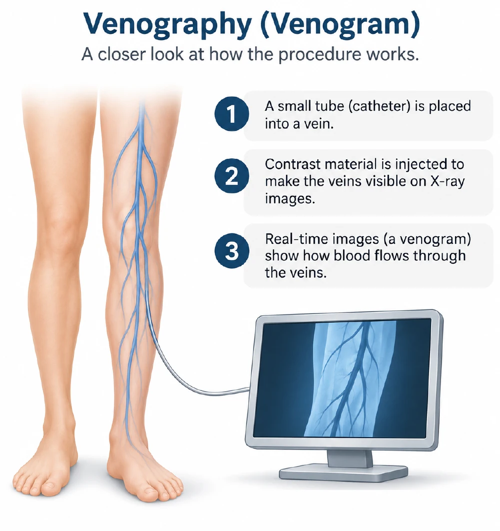
During venography, contrast material is introduced into a vein to make the blood vessels visible through X-ray imaging. This allows the physician to examine vein structure, observe blood flow, and identify possible abnormalities.
The procedure generally involves the following steps:
- Preparation: The care team reviews the patient’s medical history, medications, allergies, and any relevant previous imaging.
- Vascular access: A needle or thin catheter is placed into a vein. Local anesthetic may be used to help minimize discomfort.
- Contrast injection: Contrast material is introduced into the vein to make the blood vessels visible.
- X-ray imaging: Images are captured as the contrast moves through the venous system, allowing the physician to examine the veins.
- Image evaluation: The physician reviews the images to identify possible narrowing, blockages, or other abnormalities.
- Completion: The needle or catheter is removed, and the access site is monitored for bleeding or other concerns.
Patients may experience mild pressure near the access site or a temporary warm sensation when contrast material is introduced. The length of the procedure varies depending on which veins are being examined and whether additional vascular care is performed.
Venography vs. Venous Ultrasound
Venography and venous ultrasound both evaluate vein conditions, but they use different imaging methods.
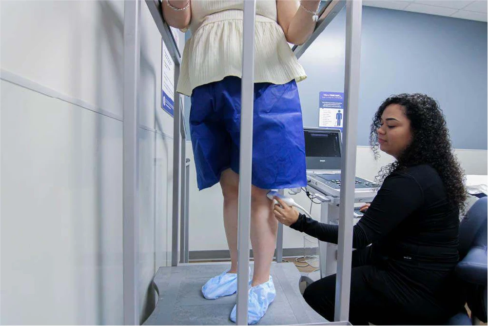
Venous ultrasound uses sound waves to examine vein structure and blood flow. It is noninvasive and does not require contrast material or radiation. Ultrasound is commonly the first imaging test used to evaluate suspected blood clots, venous reflux, and other circulation concerns.
Venography uses contrast material and X-ray imaging to provide detailed views of certain veins. It may be recommended when ultrasound findings are inconclusive or when additional imaging is needed to evaluate a suspected obstruction or plan a vascular procedure.
How Is Venography Arranged at United Vein & Vascular Centers?
A venogram may be recommended when a patient’s symptoms or previous imaging suggest a venous condition that requires further investigation. A vascular specialist will review the patient’s medical history, examination findings, and diagnostic results to determine whether additional imaging is appropriate.
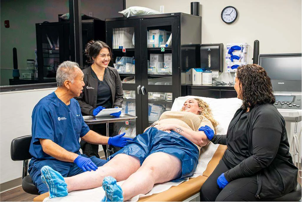
At United Vein & Vascular Centers, venography is performed at select office-based labs (OBLs). In markets where the procedure is unavailable, patients may be referred to an outside facility when their physician determines that venography is necessary.
United Vein & Vascular Centers OBLs:
Benefits and Risks of Venography
Venography offers a detailed view of the venous system that can support diagnosis and treatment planning when other imaging methods do not provide enough information.
Potential benefits include:
- Detailed visualization of vein structure and anatomy
- Identification of narrowing, blockages, or blood clots
- Additional information when previous imaging is inconclusive
- Improved planning for certain vascular procedures
- Real-time imaging guidance during selected procedures
However, venography is more invasive than ultrasound and involves exposure to contrast material and X-ray radiation.
Possible risks include:
- Bruising, bleeding, or infection at the access site
- Allergic or other reactions to contrast material
- Kidney complications related to contrast material in susceptible patients
- Blood vessel injury
- Blood clot formation
- Exposure to ionizing radiation
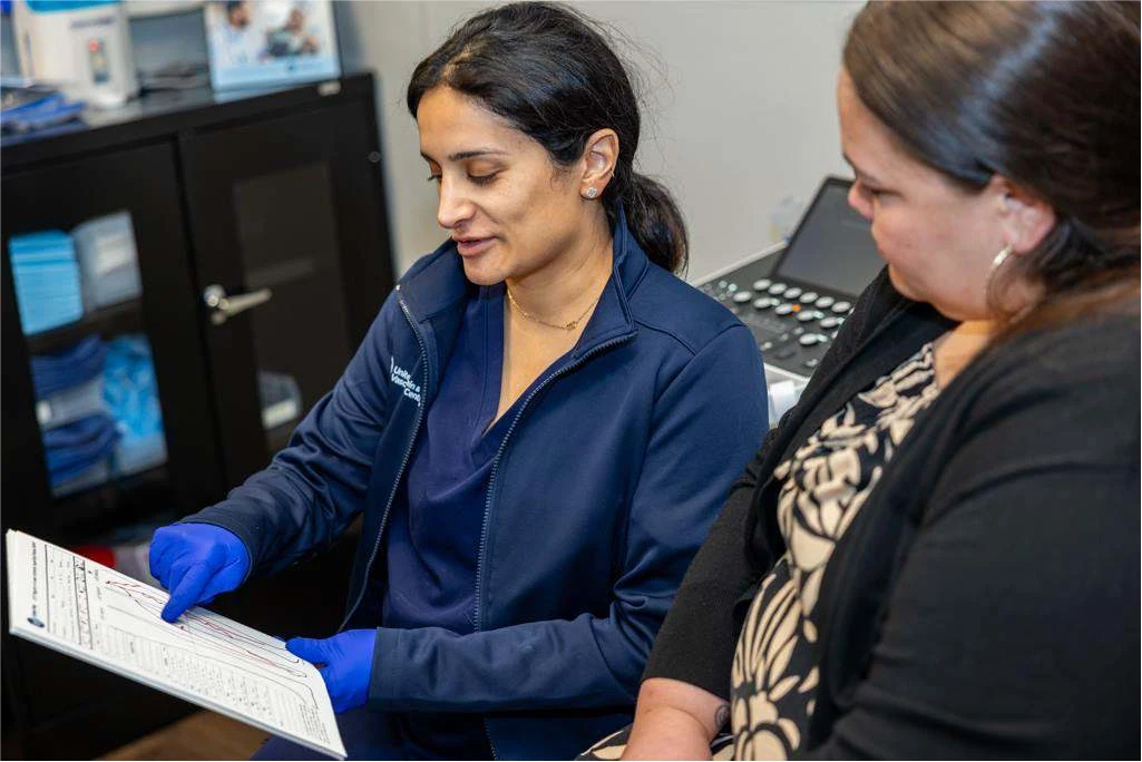
Before recommending venography, the physician will assess relevant risk factors, including kidney function, medication use, and previous reactions to contrast material. The potential benefits must outweigh the risks for the individual patient.
What Happens After Venography?
Following venography, the care team monitors the access site and provides instructions for recovery. Some patients may experience temporary tenderness or bruising where the needle or catheter was inserted.
Recovery recommendations depend on the type of venography performed and whether another procedure was completed at the same time.
After the procedure, patients may be advised to:
- Follow instructions for keeping the access site clean and protected
- Limit strenuous activity for a period recommended by their provider
- Drink fluids when medically appropriate to help clear the contrast material
- Monitor for unusual swelling, bleeding, redness, or increasing pain
- Attend any recommended follow-up appointments
The physician will review the venogram findings alongside the patient’s symptoms, medical history, and previous diagnostic testing. Depending on the results, the next step may involve continued monitoring, additional evaluation, or discussion of a minimally invasive vascular procedure.
Frequently Asked Questions
Patients may experience mild discomfort when the needle or catheter is inserted, along with temporary pressure or warmth during contrast injection. Local anesthetic may be used to help minimize discomfort.
The length of a venography varies depending on the veins being examined and whether additional imaging or vascular treatment is performed. Your care team can provide more specific expectations before the procedure.
Both procedures use contrast material and X-ray imaging to examine blood vessels. However, conventional venography focuses on the veins, while conventional angiography generally examines the arteries.
No. Venography is performed at select United Vein & Vascular Centers office-based labs (listed on the page above). If your physician determines that the procedure is necessary, but it is not available locally, you may be referred to an outside facility for testing.
Partial is invalid, missing or invalid value for property: content

Related Articles