Contents
- Medical History Review: The First Step in Diagnosing Chronic Venous Insufficiency
- Physical Exam for CVI: What Vein Specialists Look for in Your Legs
- Understanding the CEAP Classification System for Venous Disease
- Venous Duplex Ultrasound: The Gold Standard Test for CVI
- Color Doppler Ultrasound: Detecting Venous Reflux in Real Time
- How to Prepare for Your Vein Ultrasound Appointment
- How a CVI Diagnosis Guides Your Vein Treatment Plan
- Frequently Asked Questions
- Taking the First Step Toward Lighter Legs in Chicago
If your legs feel unusually heavy after a walk along the Lakefront Trail or a long shift on your feet, it may be more than everyday fatigue. Many people assume symptoms like aching, swelling, or heaviness in their legs are just part of getting older, but in many cases, they’re your body signaling an underlying circulation issue.
One common cause is Chronic Venous Insufficiency (CVI), a condition that occurs when the veins in your legs struggle to move blood back up toward your heart. Normally, tiny valves inside your veins act like one-way gates, keeping blood flowing upward. When those valves weaken or become damaged, blood can leak backward and pool in the lower legs, a process doctors call venous reflux.
The good news is that diagnosing CVI is straightforward and completely non-invasive.
Medical History Review: The First Step in Diagnosing Chronic Venous Insufficiency
The medical history review for vascular health serves as a vital investigation phase, where your specialist listens for clues that surface-level symptoms often hide. By discussing how long you have felt heaviness or throbbing, we can start determining if you are facing a temporary strain or the progression of chronic venous disease.
How you spend your working hours often dictates the pressure placed on your vascular system. Chicagoans who endure long commutes on the Kennedy or spend all day on their feet force their veins to fight gravity constantly. This sustained pressure is often the catalyst that turns a healthy vein into a damaged one, triggering common signs of poor leg circulation like evening swelling.
While lifestyle plays a major role, your family tree often holds the biggest clues. Identifying this hereditary link early helps your doctor anticipate potential complications even before they become painful.
Separating these underlying medical causes from purely cosmetic concerns sets the stage for a precise diagnosis. Once we understand the “why” behind your condition, we shift our focus to the “what.” This leads directly to the physical inspection, where we classify the visible severity of your symptoms.
Physical Exam for CVI: What Vein Specialists Look for in Your Legs
After reviewing your medical history, your vein specialist performs a visual and hands-on examination of your legs. This step helps identify visible signs of CVI and other circulation problems.
During the physical exam, your provider evaluates your legs while you are standing. Gravity naturally places pressure on the veins in your lower legs, which makes symptoms easier to detect.
Common signs of CVI doctors look for:
- Bulging varicose veins that appear twisted or enlarged
- Swelling in the ankles or calves that worsens throughout the day
- Skin texture changes such as thickening or tightening
- Pitting edema, which occurs when pressing the skin leaves a temporary indentation
- Hyperpigmentation, a reddish-brown discoloration near the ankle caused by blood leaking from damaged veins
These physical clues help determine whether poor circulation is affecting your lower legs.
However, visible symptoms only tell part of the story. To better classify the severity of venous disease, specialists use a standardized medical system.
Understanding the CEAP Classification System for Venous Disease
Vein specialists rely on the CEAP classification system to categorize the severity of chronic venous insufficiency. CEAP stands for Clinical, Etiology, Anatomy, and Pathophysiology.
This system helps providers track disease progression and determine the best treatment plan.
Think of this as a “severity score” ranging from C0 to C6 that helps track disease progression:
- C0 – C2: Ranges from no visible signs to spider veins and bulging varicose veins.
- C3: Presence of swelling (edema) in the ankles or legs.
- C4: Visible skin changes, such as brown discoloration or hardening of the skin texture.
- C5 – C6: Presence of a healed ulcer or an active, open venous leg ulcer.
While CEAP helps categorize visible symptoms, it cannot identify which veins are failing internally. For that, specialists use diagnostic imaging.
Venous Duplex Ultrasound: The Gold Standard Test for CVI
A venous duplex ultrasound is the most accurate and widely used test to diagnose chronic venous insufficiency.
This non-invasive imaging test allows specialists to see how blood flows through the veins in your legs.
The ultrasound performs two important functions:
1. Vein structure imaging
B-mode ultrasound creates detailed black and white images of your veins. These images allow the sonographer to:
- Identify previous blood clots
- Measure vein size
- Detect structural abnormalities
- Map the overall vein network in your legs
2. Blood flow evaluation
The second component evaluates how blood moves through those veins. This helps determine whether valves are functioning properly.
Many vein ultrasounds are performed while you are standing or slightly upright. Testing in this position allows gravity to reveal circulation problems that might not appear when lying flat.
Because the test is painless and requires no needles or dye, it has replaced older diagnostic procedures such as venograms.
Once the structure of the veins is mapped, the next step is measuring the direction of blood flow.
Color Doppler Ultrasound: Detecting Venous Reflux in Real Time
During the ultrasound, the technician activates color Doppler imaging to track blood movement inside the veins.
Color Doppler overlays red and blue colors onto the ultrasound image, allowing specialists to quickly see how blood is flowing.
What doctors look for during Doppler imaging:
- Normal veins show smooth upward blood flow toward the heart
- Damaged valves allow blood to flow backward toward the ankle
- Backward flow lasting more than half a second indicates venous reflux
To test how the valves respond under pressure, the sonographer may gently squeeze your calf during the scan. This mimics the pumping action that occurs when walking.
If blood flows backward after the pressure is released, the ultrasound records how long the reflux lasts. This helps identify exactly which veins are contributing to your symptoms.
The final ultrasound results provide a complete map of your circulation, allowing specialists to pinpoint the source of vein disease.
How to Prepare for Your Vein Ultrasound Appointment
Vein ultrasounds are simple outpatient procedures. A few small steps can help ensure the most accurate results.
Before your appointment:
- Drink 16 to 24 ounces of water to help veins appear more clearly on the scan
- Wear loose shorts or athletic clothing for easy leg access
- Avoid applying lotion or moisturizer to your legs that day
- Plan for a visit lasting about 45 to 60 minutes
Staying hydrated helps prevent veins from constricting, which makes them easier to evaluate during imaging.
Once your ultrasound is complete, your specialist can begin developing a targeted treatment plan.
How a CVI Diagnosis Guides Your Vein Treatment Plan
A venous ultrasound transforms vague symptoms into a precise roadmap for treatment.
For example, the bulging vein you see on your calf may actually originate from a failing vein higher in the thigh or groin. Treating that underlying vein prevents pressure from building again in the future.
A confirmed diagnosis also helps demonstrate medical necessity for insurance coverage. Ultrasound data measures vein size and reflux time, which insurers use to determine whether treatment is medically required.
Early diagnosis offers important health benefits. Treating CVI early can help prevent:
- Chronic leg swelling
- Skin thickening and discoloration
- Venous leg ulcers
- Blood clots
When circulation problems are treated sooner, patients are more likely to maintain an active lifestyle.
Frequently Asked Questions
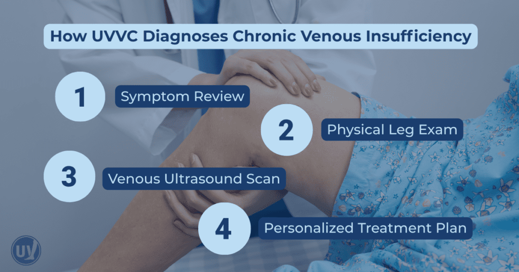
Doctors diagnose CVI through a combination of medical history review, a physical exam, and a venous duplex ultrasound that measures blood flow and detects venous reflux.
No. A venous duplex ultrasound is completely non-invasive and painless. The test uses sound waves to create images of your veins and typically takes less than an hour.
Most vein evaluations last 45 to 60 minutes. During that time, your provider reviews your symptoms, performs a physical exam, and completes a vein ultrasound.
Taking the First Step Toward Lighter Legs in Chicago
You do not have to live with heavy, swollen legs or wonder what is causing your symptoms. A simple diagnostic visit can provide clear answers and a path toward relief.
At United Vein & Vascular Centers, our Chicago specialists use advanced ultrasound technology to diagnose chronic venous insufficiency and develop personalized treatment plans.
Schedule a consultation at a UVVC Chicago clinic near you to take the first step toward healthier circulation and lighter legs.