
Understanding Vein Disease
Vein disease occurs when veins have difficulty returning blood toward the heart, most commonly affecting the legs. It often develops when valves inside the veins weaken or become damaged, allowing blood to flow backward and collect. Over time, increased pressure within the veins can contribute to leg discomfort, swelling, visible veins, skin changes, and other complications.
At United Vein & Vascular Centers, our vein specialists evaluate and treat vein disease using minimally invasive treatment options based on each patient’s symptoms, vein anatomy, ultrasound findings, and individual health needs.
Request a Vein Screening
Understanding Your Vein Health
Vein disease can affect people differently depending on which veins are involved and how well blood is circulating through the legs. Learning about symptoms, vein conditions, and available treatments can help you better understand your vein health and when an evaluation may be appropriate.
Vein Disease Symptoms
Leg swelling, aching, heaviness, cramping, itching, skin changes, and visible veins are among the symptoms that may be associated with vein disease. Because these symptoms can also have other causes, an evaluation can help determine whether venous circulation may be contributing to your concerns.
Choroby żył
Problems with blood flow and increased pressure within the veins can contribute to conditions including chronic venous insufficiency, varicose veins, reticular veins, spider veins, venous stasis dermatitis, and venous ulcers.
Zabiegi na żyły
When treatment is appropriate, minimally invasive procedures can be used to close or remove affected veins so blood can naturally reroute through healthier veins.
How Do the Veins in the Legs Work?
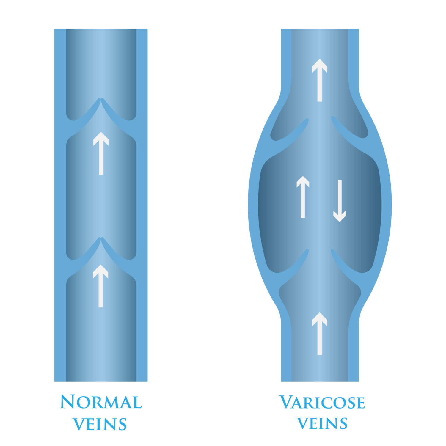
Veins return blood from the body to the heart. Because veins in the legs must move blood upward against gravity, they rely on small, one-way valves that help prevent blood from flowing backward. The calf muscles also help by squeezing the veins during movement and pushing blood out of the lower legs.
The veins in the legs form an interconnected network made up of deep, superficial, and perforator veins.
Deep Veins
Deep veins are located within or alongside the leg muscles and carry most of the blood from the legs toward the heart. Because they perform an essential role in circulation, minimally invasive treatments for superficial vein disease generally do not target healthy deep veins.
A blood clot that forms in a deep vein is called deep vein thrombosis (DVT). DVT is different from chronic superficial vein disease and requires prompt medical evaluation. Sudden pain, swelling, warmth, or redness, particularly in one leg, should not be ignored.
Superficial Veins
Superficial veins are located closer to the skin and collect blood from the skin and surrounding tissues before directing it into the deep venous system.
The great saphenous vein and small saphenous vein are two of the major superficial veins in the legs. When valves within superficial veins do not function properly, increased pressure can contribute to visible veins and other signs of vein disease.
Perforator Veins
Perforator veins connect the superficial veins with the deep venous system. Their valves normally help direct blood inward toward the deep veins.
When perforator vein valves do not function properly, blood may flow back toward the surface, increasing pressure in nearby veins and potentially contributing to swelling, varicose veins, skin changes, or venous ulcers.
How Does Vein Disease Develop?
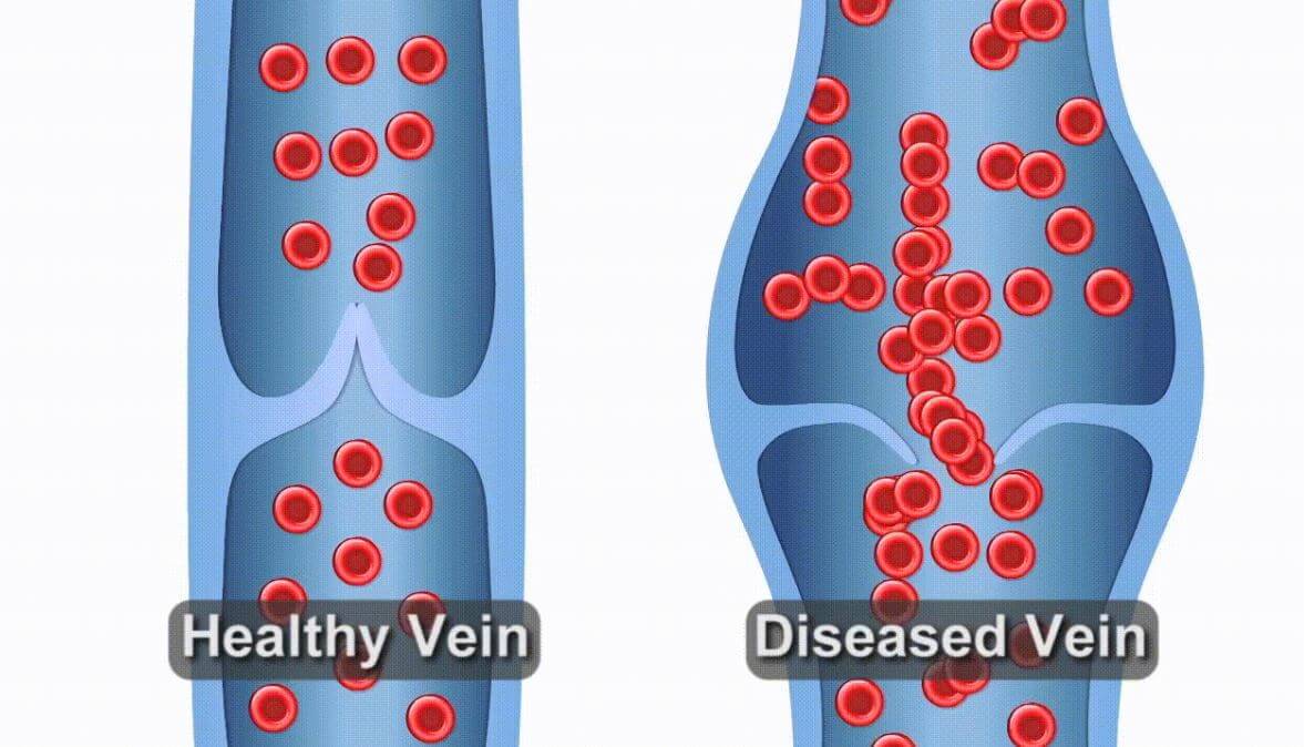
Healthy vein valves help keep blood moving in one direction toward the heart. When these valves weaken or become damaged, blood can flow backward and collect in the legs. This backward flow is known as venous reflux.
Reflux can affect different parts of the venous system. As blood continues to collect, pressure within the veins can increase.
When the veins have ongoing difficulty returning blood efficiently toward the heart, chronic venous insufficiency (CVI) may develop. Over time, increased venous pressure can contribute to persistent swelling, aching, heaviness, visible veins, and changes to the skin and surrounding tissue.
Symptoms may become more noticeable after prolonged sitting or standing because the veins must work against gravity to move blood out of the lower legs.
What Are the Most Common Vein Conditions?
Vein disease can appear in different ways depending on the veins involved, valve function, and the amount of pressure within the venous system.
Przewlekła niewydolność żylna
Chronic venous insufficiency occurs when the veins have ongoing difficulty returning blood efficiently from the legs toward the heart. CVI may contribute to swelling, aching, heaviness, skin changes, varicose veins, and other complications.
Learn About Chronic Venous Insufficiency
Żylaki
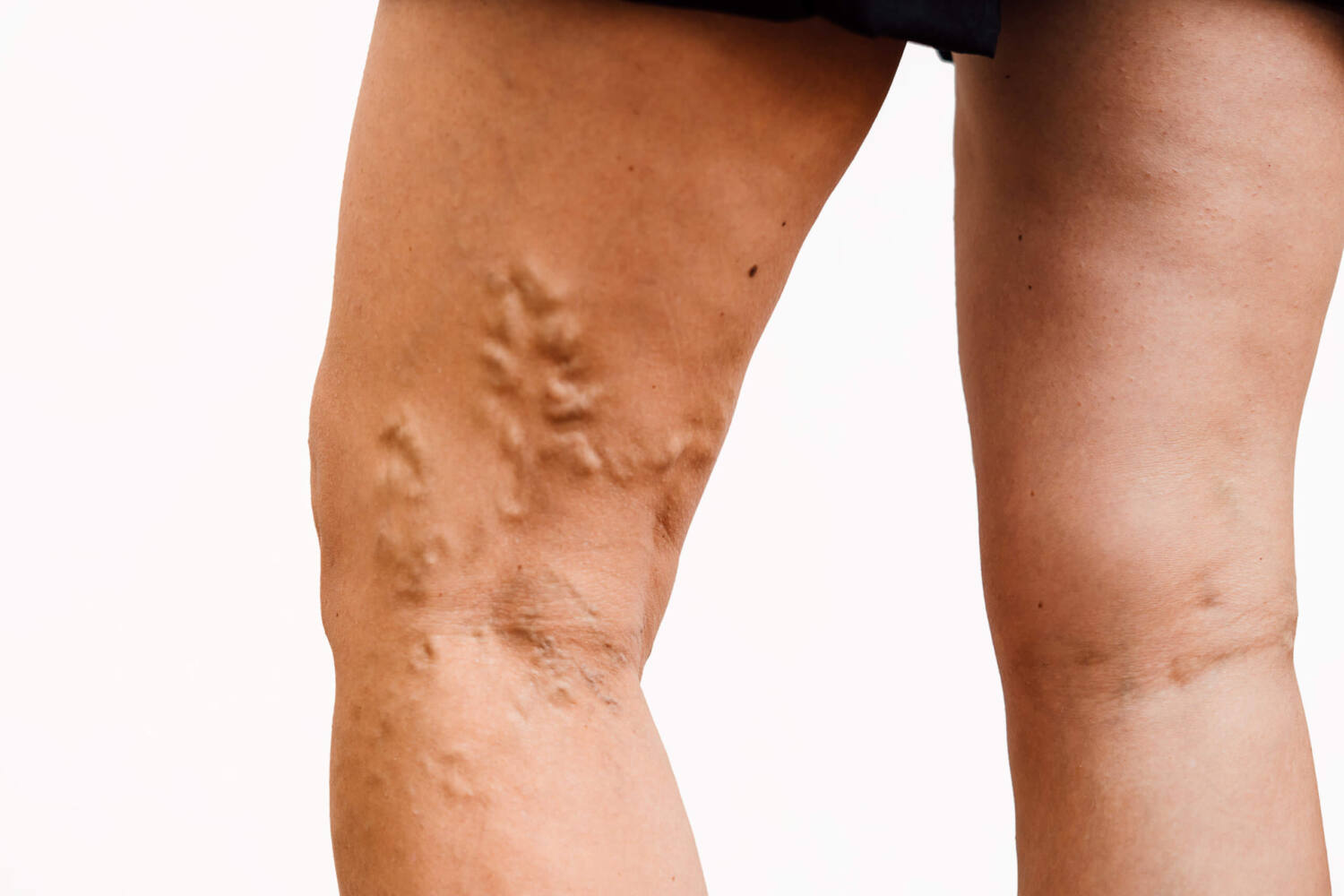
Varicose veins are enlarged, twisted veins that commonly develop in the legs. They may appear raised or rope-like and can be associated with aching, swelling, heaviness, itching, or leg fatigue.
Reticular Veins
Reticular veins are blue or green veins visible beneath the skin. They are generally larger than spider veins but smaller and flatter than varicose veins. Some patients may experience aching, tenderness, or burning around the affected area.
Pajączki
Spider veins are small red, blue, or purple vessels that often appear as thin lines, branches, or web-like patterns near the skin’s surface.
Although appearance may be the first concern, spider veins can occur alongside symptoms or other signs of vein disease. United Vein & Vascular Centers does not treat spider veins for cosmetic purposes alone. A vein screening can help determine whether visible veins may be associated with an underlying vein condition.
Venous Stasis Dermatitis
Venous stasis dermatitis is skin inflammation associated with ongoing venous insufficiency. It may cause itching, dryness, swelling, thickened skin, or reddish-brown discoloration, particularly around the lower legs and ankles.
Learn About Venous Stasis Dermatitis
Venous Ulcers
Venous ulcers are slow-healing wounds that commonly develop near the ankle when prolonged venous pressure damages the skin and surrounding tissue.
United Vein & Vascular Centers evaluates and treats vein disease that may be contributing to a venous ulcer but does not provide wound-care services.

What Increases the Risk of Vein Disease?
Several factors may increase the likelihood of developing venous reflux or chronic venous insufficiency, including:
- A family history of vein disease
- Starzenie się
- Pregnancy or hormonal changes
- Excess weight
- Długotrwałe siedzenie lub stanie
- Limited mobility
- Previous blood clots
- Prior leg injury, trauma, or surgery
- Palenie
Having one or more risk factors does not mean someone has vein disease. An evaluation is needed to determine whether symptoms or visible vein changes may be related to venous circulation.
How Is Vein Disease Evaluated?
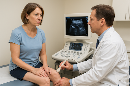
A vein screening is an initial evaluation used to review symptoms, relevant health history, and visible signs that may be related to vein disease with a board-certified vein specialist. Based on the findings, your specialist may recommend additional testing.
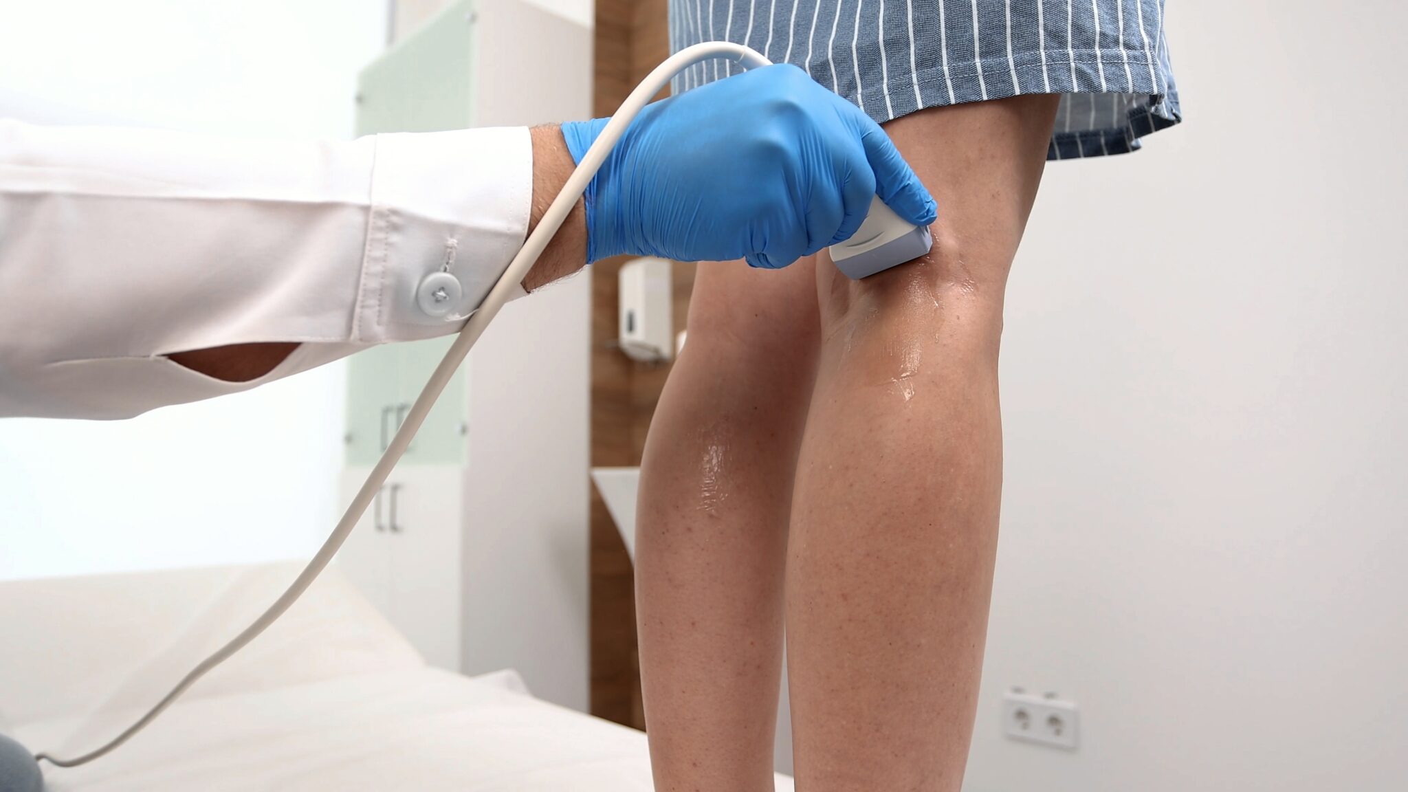
When appropriate, a duplex ultrasound can evaluate blood flow, valve function, and the deep, superficial, and perforator vein systems. It can also help identify the location and pattern of venous reflux.
A vein screening does not automatically include an ultrasound, and not every patient who completes a screening will need testing or treatment. Recommendations depend on the individual evaluation.
How Is Vein Disease Treated?
When treatment is appropriate, the recommended procedure depends on the affected veins, symptoms, medical history, ultrasound findings, and provider evaluation.
Minimally invasive vein procedures offered at United Vein & Vascular Centers may include:
Ablacja prądem o częstotliwości radiowej (RFA)
Radiofrequency ablation uses controlled thermal energy to close a targeted vein that is not circulating blood properly.
Learn About Radiofrequency Ablation
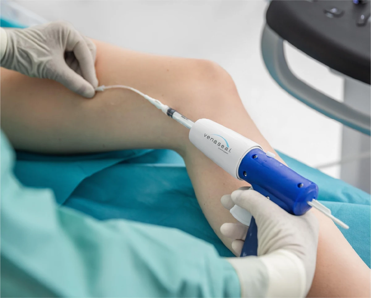
VenaSeal™
VenaSeal uses a medical adhesive to close an affected vein without the use of thermal energy.
Varithena®
Varithena is a prescription injectable microfoam used to close certain affected veins.
Skleroterapia pod kontrolą USG
Ultrasound-guided sclerotherapy uses ultrasound imaging to guide an injectable medication into a targeted vein.
Mikroflebektomia
Microphlebectomy, also known as stab or ambulatory phlebectomy, removes selected bulging varicose veins through very small openings in the skin.
Veins selected for treatment are already functioning poorly. Closing or removing an affected superficial vein allows blood to naturally reroute through healthier veins that can carry it more effectively.
Recovery varies based on the procedure and individual patient factors. Many patients return to normal daily activities relatively quickly, and walking is often encouraged after treatment. The care team provides instructions based on the procedure performed.
Często zadawane pytania
Układ krążenia odpowiada za transport krwi w całym organizmie. Tętnice przenoszą bogatą w tlen krew z serca, podczas gdy żyły odprowadzają ją z powrotem. Kiedy maleńkie zastawki wewnątrz żył słabną lub zawodzą, krew może płynąć do tyłu i gromadzić się w nogach - stan znany jako refluks żylny lub przewlekła niewydolność żylna (CVI). To słabe krążenie często prowadzi do żylaków, obrzęku nóg i dyskomfortu.
Niewydolność żylna występuje, gdy żyły nóg nie mogą skutecznie przesyłać krwi z powrotem do serca. Często jest ona niedostatecznie diagnozowana, ponieważ objawy takie jak ból, obrzęk lub widoczne żyły mogą być mylone z kwestiami kosmetycznymi lub związanymi ze starzeniem się. Bez leczenia niewydolność żylna może postępować, prowadząc do przebarwień skóry, owrzodzeń i przewlekłego bólu. Wczesna ocena przez specjalistę od żył może zapobiec powikłaniom i poprawić krążenie.
W United Vein & Vascular Centers edukacja pacjentów jest podstawą naszej opieki. Nasi certyfikowani specjaliści wykorzystują diagnostyczne obrazowanie ultrasonograficzne, aby wskazać przyczynę objawów, a następnie szczegółowo wyjaśnić wyniki i spersonalizowany plan leczenia. Oferujemy minimalnie inwazyjne zabiegi w gabinecie, które przywracają zdrowy przepływ krwi przy niewielkim lub żadnym przestoju - pomagając szybciej wrócić do ulubionych zajęć.