Contenido
- A Connected System Beneath the Skin
- Truncal Veins: The Major Pathways
- Tributaries: The Branches Patients Often Notice First
- Vein Valves and the Calf Muscle Pump
- Why the Vein You See May Not Be the Source
- How Duplex Ultrasound Creates a Venous Map
- Matching Treatment to the Affected Pathway
- The Ultrasound Shows the Pattern. The Patient Gives It Meaning.
- What I Want Patients to Understand About Their Veins
- Looking at the Whole System
By Domenic Zambuto, M.D., RPVI, DABVLM
Patients usually begin by showing me what they can see. A raised vein crosses the calf; a cluster of veins has appeared near the knee. One ankle looks swollen by the end of the day, or the skin around it has slowly changed color.
Those observations matter. They tell us where a patient is noticing a problem. But they do not always tell us where that problem began.
The veins in the legs are not isolated vessels. They form a connected network, with larger veins receiving blood from smaller branches and other veins linking pathways at different depths. Valves inside those vessels work with the calf muscles to move blood upward against gravity. When part of that system stops working effectively, the effects can appear farther along the network.
That is why thoughtful vein care starts by looking beyond the most visible vein. We need to understand the structure beneath the skin, the direction in which blood is moving, and the relationship between a patient’s symptoms and the anatomy involved.
A Connected System Beneath the Skin
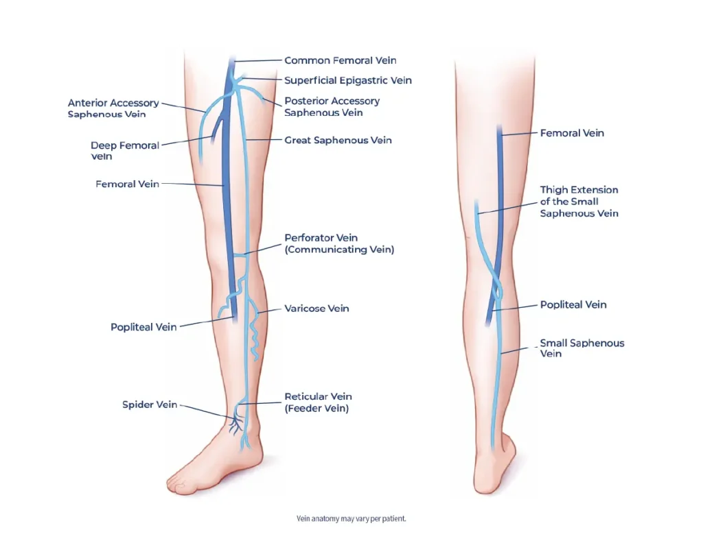
The veins in the legs can be organized into three connected groups: the deep venous system, the superficial venous system, and the perforator veins that run between them.
Deep veins lie within the muscles and carry most of the blood returning from the legs toward the heart. Movement plays an important role in that process. When the calf muscles contract during walking, they compress the deep veins and help propel blood upward.
The superficial venous system is located closer to the skin. It gathers blood from the surrounding tissues and directs it toward the deep system. Perforator veins connect the two. When they are functioning properly, their valves help keep blood moving from the surface of the leg toward the deeper pathways.
Each group has a distinct role, but none works alone. A change in pressure or valve function within one pathway may affect other veins connected to it. To make sense of what we see on the outside of the leg, we first have to understand those relationships underneath it.
Truncal Veins: The Major Pathways
Within the superficial venous system, the larger longitudinal pathways are known as truncal veins. The three most familiar are the great saphenous vein, the small saphenous vein, and the anterior saphenous vein.
The great saphenous vein, or GSV, begins near the foot and travels along the inner side of the leg and thigh before joining the deep venous system near the groin. Because of its length and the number of branches that drain into it, the GSV is an important part of many venous evaluations.
The small saphenous vein generally travels up the back of the calf and commonly joins the deep system behind the knee. Its course and connections vary, however, and that variation can influence how reflux develops and how treatment is planned.
The anterior saphenous vein follows its own path along the front portion of the thigh. It was once commonly called the anterior accessory saphenous vein, but current terminology recognizes it as a truncal vein in its own right. That distinction is more than a matter of vocabulary. Accurate names help physicians describe the anatomy clearly and communicate exactly which pathway is involved.
The term “truncal” comes from the word “trunk.” Much like the trunk of a tree supports and receives smaller branches, truncal veins are the larger superficial veins into which smaller tributary veins drain. The great saphenous, small saphenous, and anterior saphenous veins serve as these primary pathways, carrying blood toward connections with the deep venous system.
Tributaries: The Branches Patients Often Notice First
Tributaries are smaller veins that drain into the truncal veins. When pressure increases within the venous system, these branches may enlarge, twist, or become visible beneath the skin. Many veins that patients recognize as varicose veins are tributaries.
This is where appearance can be misleading.
A bulging tributary may be the most obvious finding on the leg, but the abnormal pressure affecting it may originate in a connected truncal vein. Treating only the branch that can be seen, without first determining what is feeding it, may leave an important part of the pattern unexplained.
I sometimes describe the system as a network of main roads and side streets. The truncal veins are the larger routes, tributaries feed into them, and perforator veins create connections between the superficial and deep systems. If flow becomes abnormal along one route, the pressure may be felt elsewhere in the network.
It is a simple comparison, but it captures an important clinical principle: location is not the same as source.
Vein Valves and the Calf Muscle Pump
The structure of a vein matters, but so does what is happening inside it.
Unlike arteries, which carry blood away from the heart under higher pressure, leg veins must return blood upward while working against gravity. They rely on movement and a series of small, one-way valves to do that efficiently.
As the calf muscles contract, they squeeze the veins and move blood upward. The valves then close to help limit backward flow between muscle contractions. This partnership is often called the calf muscle pump.
When the valves do not close effectively, some blood can move backward. That backward flow is known as venous reflux. It can raise pressure within the affected pathway and place additional strain on connected veins.
Over time, patients may develop symptoms or visible changes such as:
- Aching, throbbing, or cramping in the legs
- Heaviness or fatigue that builds during the day
- Swelling around the ankles or lower legs
- Itching or burning
- Restlessness in the legs
- Enlarged or twisted varicose veins
- Skin discoloration or changes in texture
- Hard-to-heal leg ulcers
There is no single appearance that defines venous reflux. One patient may have prominent varicose veins with relatively little discomfort. Another may experience daily aching, heaviness, or swelling without many visible veins. The symptoms, examination, and blood-flow findings all have to be considered together.

Why the Vein You See May Not Be the Source
When a patient points to a vein along the calf, I pay attention to its location and pattern. Then I begin asking a different set of questions.
Where is that branch receiving blood? Are the valves in the connected truncal vein working properly? Does backward flow begin near the groin, behind the knee, within a shorter segment, or somewhere else? Are perforator veins contributing to the pressure? Is the deep venous system functioning as expected?
The answers cannot be determined by appearance alone.
Venous anatomy varies from one person to another. Reflux may affect the great saphenous vein, small saphenous vein, anterior saphenous vein, tributaries, perforator veins, or more than one connected pathway. Even two patients with varicose veins in a similar location may have different patterns beneath the skin.
The goal is not to make the anatomy more complicated than it needs to be. It is to avoid oversimplifying the problem before we understand it.
How Duplex Ultrasound Creates a Venous Map
When clinically appropriate, duplex ultrasound allows the care team to examine the structure of the veins and the movement of blood through them. The study combines traditional ultrasound imaging, which shows the veins and surrounding anatomy, with Doppler technology, which evaluates the direction and speed of blood flow. Together, these components provide information that a visual examination alone cannot reveal.
During an ultrasound evaluation, we can assess whether the veins are open, observe the direction of blood flow, and examine how well the valves are functioning. We can identify where reflux begins, follow how far it extends, and see which truncal veins, tributaries, or perforator veins are involved. The deep venous system is evaluated as part of that larger picture.
In practical terms, the study creates a map. That map helps connect a patient’s symptoms with a specific pattern of blood flow. It also helps us avoid assuming that the vein drawing the most attention is automatically the vein that should be treated first—or treated at all.
Ultrasound does not replace the conversation with the patient. It gives that conversation anatomical context.
Matching Treatment to the Affected Pathway
Once we understand the symptoms, examination findings, medical history, and ultrasound results, we can determine whether treatment may be appropriate and which option fits the affected anatomy.
If reflux is present within a truncal vein, care may focus on closing the diseased section so blood can redirect through functioning veins. If enlarged tributaries remain symptomatic or clinically significant, they may require a different approach. Some patients have more than one area that needs to be addressed over the course of care.
At United Vein & Vascular Centers, minimally invasive treatment options may include:
| Treatment | How it works |
| Ablación por radiofrecuencia | Uses controlled thermal energy to close an affected vein. |
| VenaSeal | Uses a medical adhesive to close a diseased venous pathway. |
| Varithena | Uses a prescription injectable microfoam to treat certain incompetent veins. |
| Ultrasound-guided sclerotherapy | Uses ultrasound to guide an injectable medication into targeted veins. |
| Microflebectomía | Removes selected varicose tributaries through very small openings in the skin. |
These procedures are not interchangeable, and a patient does not necessarily need every option listed. A treatment used for a refluxing truncal vein may not be the same treatment used for a visible tributary. Recommendations depend on the location and extent of reflux, the size and course of the veins, the patient’s symptoms, medical history, and other individual considerations.
Patients sometimes ask how blood can still leave the leg after a diseased vein is closed. The answer goes back to the network itself. When an affected vein is appropriately treated, blood can redirect through other functioning veins that continue carrying it toward the heart. We are not removing the body’s ability to circulate blood; we are addressing a pathway that is no longer moving it efficiently.
The Ultrasound Shows the Pattern. The Patient Gives It Meaning.
An anatomical map can show me where reflux is present. It cannot tell me that swelling makes a patient’s shoes feel tight at the end of a shift. It cannot explain that leg heaviness has shortened an evening walk or that aching regularly interrupts sleep.
Those details matter because similar ultrasound findings do not always affect people in the same way. One patient may be limited by discomfort and swelling. Another may be most concerned about recurring skin changes. Someone else may have visible varicose veins but few physical symptoms.
Good care requires both forms of information. The ultrasound helps us understand what blood is doing beneath the skin. The patient tells us how the condition affects everyday life.
We should not treat an image in isolation. We should use that image, together with the patient’s experience and clinical findings, to decide whether care is needed and what a reasonable plan may look like.
What I Want Patients to Understand About Their Veins
If patients remember only a few points, I hope they are these:
- The veins in the legs form a connected network rather than a collection of isolated vessels.
- The great saphenous, small saphenous, and anterior saphenous veins are major superficial truncal pathways.
- Tributaries are smaller branches that drain into larger veins and may become visibly varicose.
- Perforator veins connect the superficial and deep venous systems.
- Vein valves and the calf muscles work together to move blood upward.
- When valves do not close effectively, venous reflux can raise pressure within connected veins.
- The most visible vein may not be the original source of the problem.
Looking at the Whole System
After more than two decades of diagnosing and treating chronic venous insufficiency, I continue to return to the same principle: the quality of a treatment plan depends on the quality of the evaluation behind it.
At United Vein & Vascular Centers, we look at the patient’s symptoms, examine the legs, and use ultrasound when clinically appropriate to understand what is happening beneath the surface. From there, the care team can determine whether venous disease may be contributing to the patient’s concerns and recommend care based on confirmed findings.
That process is more thoughtful than chasing individual veins as they appear. It recognizes that a raised tributary, a refluxing truncal vein, a connecting perforator, and the valves within them are all parts of the same working system.
When patients understand that system, the treatment conversation becomes clearer. They can see why the area that hurts or bulges may not be where the problem started and why mapping the entire pathway is an essential part of deciding how UVVC may be able to help.